| |
PATHOPHYSIOLOGY OF CO2 EXCHANGES
1. Physiology of CO2 production and excretion
CO2 is produced by the metabolism and eliminated (mainly) via respiration at a rate of approx. 8-10 mmol/min. At a blood flow of 5 L/min. this implies that each liter of blood uploads 1.6-2 mmol of CO2 in the systemic capillaries and releases the same amount of CO2 in the pulmonary capillaries.
The blood contains 20-22 mmol/L of CO2 (if measurement is carried out in the plasma 24-26 mmol/L), in three forms:
| dissolved CO2 | bicarbonate (HCO3-) | protein bound carbamates |
| 1.2 mM (40 mmHg) | 24 mM (plasma) 18 mM (whole blood) | 1.2 mM |
| 5% | 90% (plasma) | 5% |
The solubility of CO2 is only 0.03 mM/mmHg at 37o C, and in water, at a partial pressure of 40 mmHg, little bicarbonate would be formed and the total CO2 content would be only slightly higher than 1.2 mM. The very high concentration of total CO2 in blood is achieved because of the presence of other buffers (hemoglobin, serum albumin, phosphate) that maintain pH=7.4 and promote the dissociation of CO2 to bicarbonate, a non-volatile component that has no effect on PCO2. The reaction is as follows:
CO2 + X + H2O ↔ HCO3- + HX+ (eq. 1)
where X represents the non-carbonic buffers.
At the level of the tissues most CO2 is converted to bicarbonate, thanks to the action of carbonic anhydrase, whereas the opposite occurs at the level of the lung capillaries. It is important to remark that the amount of CO2 eliminated by the lungs (8-10 mmol/min., corresponding to 1.6-2 mM/L of blood) exceeds the CO2 content and the process could not function in the absence of the non-carbonic buffers and reversible conversion to bicarbonate.
The concentration of non-carbonic buffers can be estimated from the cumulative buffer capacity of whole blood, which ranges between 31 and 38 mEq/L; of this value the couple CO2/bicarbonate only accounts for 4-5 mEq/L, the remaining 26-33 mEq/L representing the cumulative buffer capacity of non-carbonic buffers.
Eq. 1 shows that the sum of deprotonated buffer components (Sdp) of blood, under physiological conditions is constant, because we can deprotonate CO2 only at the expense of protonation of non-carbonic buffers and vice versa.
We can write down a mathematical model of CO2 transport under the assumption that the behaviour of the non carbonic buffers (X) can be described as that of a single buffer component with acidic constant KX and concentration CX = [HX+] + [X]; this approximation is valid as long as small pH changes are considered. The model is as follows:
Sdp = [X] + [HCO3-]
[H3O+] = KC x 0.03 x PCO2 / [HCO3-] = KX x [HX+] / [X]
We observe that:
[HCO3-] = Sdp - [X]
[HX+] = CX - [X]
and we obtain:
KC x 0.03 x PCO2 / (Sdp - [X]) = KX x (CX - [X]) / [X]
yielding:
[X]2 - [X] (Sdp + CX + 0.03 x PCO2 x KC/KX) + Sdp x CX = 0
Once the above equation is solved for [X], one can calculate all other parameters of the blood gas analysis (e.g. [HCO3-], pH, standard pH, standard bicarbonate and base excess).
A problem of this model, beyond the approximation of using one single component (X) for many, is that the terms Sdp, CX, and KX are dummy parameters that do not correspond to any precisely measurable quantity; however one can reasonably fix KX at 10-7 M, and derive the other two terms from pH and the buffer capacity of the blood, either directly measured or estimated from the concentrations of hemoglobin, serum albumin and phosphate. The scope of the model is that of taking three parameters obtained from the blood gas analysis (pH, PCO2, and either the buffer capacity of blood or the concentration of hemoglobin, from which CX and Sdp can be derived) and determine the pH changes induced by varying PCO2, e.g. to simulate venous blood or acute respiratory acidosis. This model is implemented at this link.
2. Other acids and bases
The diet and metabolism under physiological and pathological conditions introduce or produce some bases and acids other than CO2 that the lung cannot eliminate, and must be dealt with by the kidneys. These are:
- (i) ammonia derived from the deamination of alimentary aminoacids; the amount depends on the diet but may be estimated at 700-1,000 mmol/day. Ammonia is almost completely protonated to ammonium ion and then converted to the non-basic urea or to glutamine.
- (ii) sulfuric acid (H2SO4) derived from the catabolism of Cys and Met; the amount depends on diet but an average 30 mEq/day can be estimated;
- (iii) phosphate, derived from the diet;
- (iv) other organic acids and their salts.
The net dietary balance of acids and bases may be basic or acidic; on average it is verges on the acidic and introduces approx. 50 mEq /day of non-volatile acids in our body.
The kidneys offer the second fundamental contribution to pH homeostasis, by excreting acids or bases in the urine. Strictly speaking, the urine is not a buffered solution; its pH ranges between 4.5 and 8 and is mainly determined by the hydrolysis of either of two ionic components: ammonium ion and citrate. The only ion in the urine that can behave as a buffer, at least under some conditions, is phosphate. The hydrolysis of citrate is basic, that of ammonium is acidic. Ammonium ion is not filtered by the kidney (its concentration in the serum being very low, at < 50 μmol/L), but is produced by the proximal tubule by deamination of glutamine (catalyzed by glutaminase) and glutamic acid (catalyzed by glutamate dehydrogenase). The end products are ammonia, which is rapidly protonated to ammonium ion, and α-ketoglutarate, which is converted to oxalacetate and then to PEP, with release of two molecules of CO2. The overall reaction is as follows:
C5H10O3N2 + 3 H2O ↔ C3H6O3 + 2 HCO3- + 2 NH4+ (eq. 2)
where lactate, bicarbonate and approximately 50% of the ammonium produced are reabsorbed in the blood, whereas the remaininng 50% of ammonium is excreted. The reabsorbed ammonium is rapidly converted to urea by the urea cycle in the liver. The overall overall balance of the urea cycle is:
2 HCO3- + 2 NH4+ ↔ CO(NH2)2 + CO2 + 3 H2O (eq. 3)
thus, the urea cycle consumes equimolar amounts of ammonia and bicarbonate and produces equimolar amounts of CO2 and urea.
The average amount of ammonia excreted in the urine is 30-50 mg/day, but it can increase to 200 mg/day in the course of metabolic acidosis. Protonation of ammonia in the urine is essentially complete and requires proton donation from CO2 (eq. 2) or from the blood buffers; thus, urinary excretion of ammonia effectively raises the blood pH and increases the fraction of deprotonated components of blood buffers (see figure below). Phosphate, citrate and other ions are secreted by kidney cells or filtered from the blood and reabsorbed to a variable degree depending on the blood and urine pH.
The kidney eliminates relatively little acid compared to the lung (respectively up to 200 mEq/day vs. 15 mol CO2/day) but has a fundamental advantage: it can select with its pumps which ions are to be eliminated and which are to be reabsorbed. Control over these processes is exerted by pH itself and by hormones. Consistently the urine pH can vary from 4.5 to 8, and the kidney can alter the sum of deprotonated components of blood buffers, a result that the lung cannot achieve.
3. Respiratory acidoses
If the CO2 exchange capability of the lungs is suddenly decreased by a disease (type 2, hypercapnic respiratory failure, e.g. because of hypoventilation in a comatous state), the PCO2 gradient required to eliminate the CO2 produced by metabolism increases, and the blood gas analysis registers an increased arterial PCO2. This phenomenon if called acute respiratory acidosis and has three hallmarks: (i) a significant increase of PCO2; (ii) a significant decrease of blood pH; and (iii) a moderate increase of bicarbonate. The pH decrease and bicarbonate increase induced by a given change in PCO2 can be calculated using the equation described in the exercise on blood buffers. The reason of the small increase of bicarbonate concentration in acute respiratory acidosis is that formation of bicarbonate requires proton donation by the non-carbonic buffers (eq. 1) and is limited by the fact that the sum of deprotonated buffer components is constant.
If the patient survives and the pulmonary condition is not resolved, the kidneys compensate for the pH imbalance by producing and reabsorbing bicarbonate. The reaction requires production of ammonia from glutamine (eq. 2). Ammonium ion is excreted in the urine (together with chloride), whereas bicarbonate (together with sodium) is reabsorbed in the blood. The urine becomes more acidic because of the hydrolysis reaction of ammonium, and excretion of hydrogen ions is effectively increased.
The kidneys increase the bicarbonate concentration in the blood bypassing reaction 1; thus in this process the sum of deprotonated buffer components of blood is effectively increased, and the condition of the patient evolves to that of a chronic compensated respiratory acidosis, whose hallmarks are: (i) strongly increased PCO2; (ii) strongly increased bicarbonate; and (iii) moderately decreased pH. The effect of bicarbonate on pH is defined by Henderson and Hasselbalch equation:
pH = pK + log ([HCO3-] / 0.03xPCO2) (eq. 4)
A scheme of the chemical reactions occurring in the blood, lungs and kidneys is as follows:
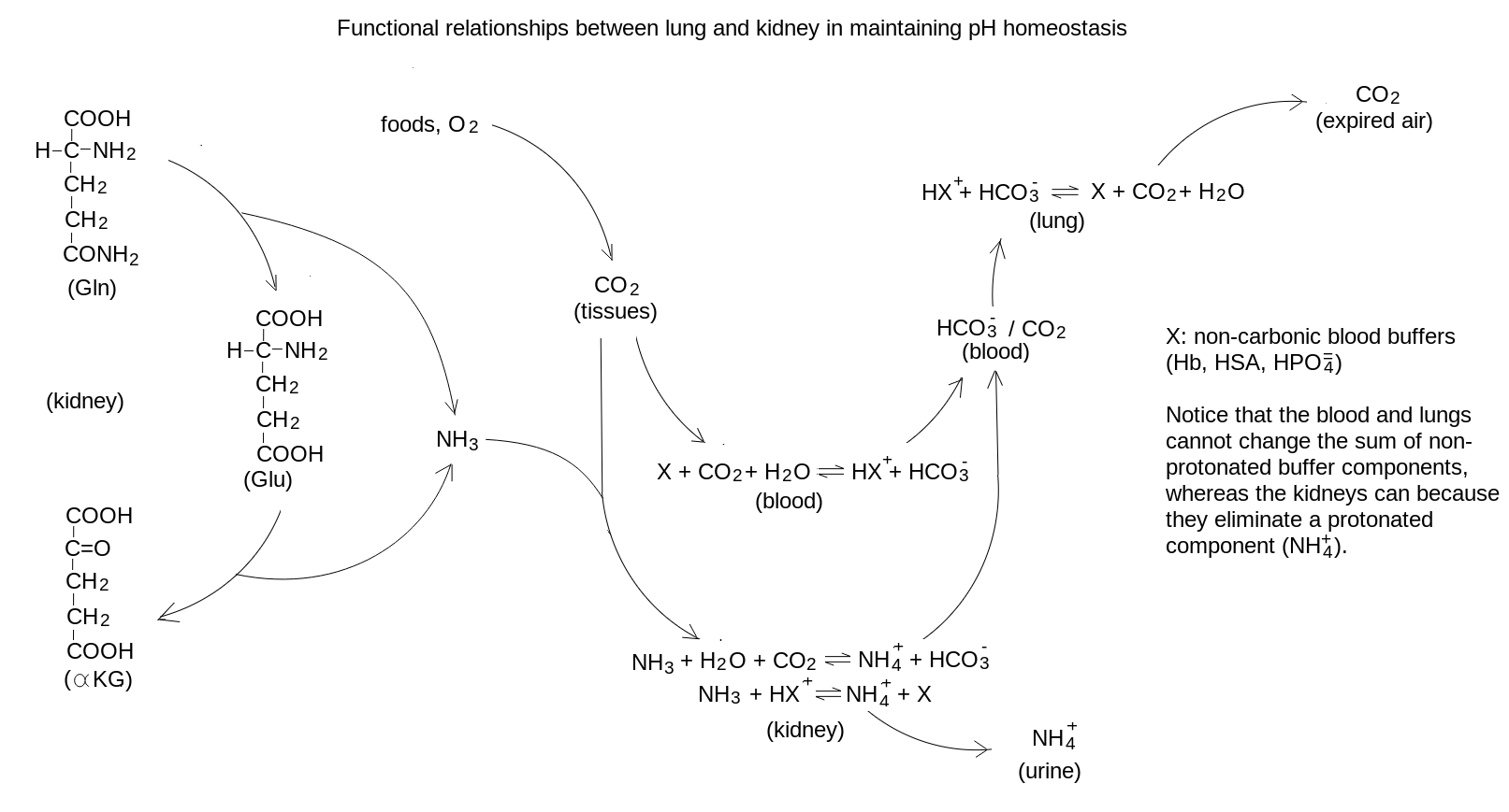 Since the lungs excrete approx. 15 moles of CO2 per day, whereas the kidneys cannot excrete more than 200 mmoles of NH4+ per day (normal value30-50 mmoles per day, but significant increases are possible in the course of extra-renal acidoses), full compensation of respiratory acidoses requires several days (up to 1 week). The expected optimal compensation can be calculated using Winter's empyrical formulas.
Typical results of a blood gas analysis of respiratory acidoses, as compared with the healthy conditions, are as follows:
| Table 1:examples of blood gas parameters for respiratory acidoses |
| | healthy condition | acute respiratory acidosis | chronic respiratory acidosis |
| arterial pH | 7.4 | 7.24 | 7.34 |
| arterial PCO2 | 40 mmHg | 70 mmHg | 70 mmHg |
| arterial HCO3- | 22.7 mM | 29.2 mM | 36.5 mM |
| total CO2 | 23.9 mM | 31.3 mM | 38.6 mM |
| buffer capacity of blood | 38.6 mEq/L | 47.7 mEq/L | 42.7 mEq/L |
| sum of deprotonated buffer components | 81 mEq/L | 81 mEq/L | 89.3 mEq/L |
| Base excess of blood | - | - | 10.3 mEq/L |
Comments to table 1: (i) in acute respiratory acidosis some parameters are identical or close to those of the healthy condition: e.g. BE is zero in both conditions, and total CO2 is increased by less than 20%. However, PCO2 is significantly increased and pH is significantly decreased. The changes in the sum of deprotonated buffer components and buffer capacity are attributed to the change in pH. (ii) In chronic respiratory acidosis the pH change with respect to the healthy condition may be minimal, but other parameters are grossly altered, e.g. bicarbonate and total CO2 are increased by more than 60%. The increase of bicarbonate is due to reaction 2, and is the cause of the increased sum of the deprotonated buffer components, and the large base excess.
4. Respiratory alkaloses
Hyperventilation causes the arterial PCO2 to move in the direction of the PCO2 of inspired air, which is very low (usually less than 0.1% or 0.7 mmHg). Causes of hyperventilation may be anemia, adaptation to high altitude, neurological lesions, etc. If the disturbance occurs acutely, we observe acute respiratory alkalosis, characterized by low arterial PCO2, significnatly increased pH and moderately reduced bicarbonate. The reasons for these changes are similar to those considered for acute respiratory acidosis: the decrease of CO2 exceeds that of bicarbonate, and is achieved only via reaction 1; this limits the respiratory loss of bicarbonate. Over a time course of a few days, however, renal compensation occurs: production and excretion of ammonia is reduced, while urinary loss of bicarbonate is increased. As a consequence the concentration of bicarbonate is reduced, together with the sum of deprotonated buffer components. The decrease of bicarbonate tends to restore the pH according to eq. 3. Typical results of a blood gas analysis of respiratory alkaloses, as compared with the healthy conditions, are as follows:
| Table 2: examples of blood gas parameters for respiratory alkaloses |
| | healthy condition | acute respiratory alkalosis | chronic respiratory alkalosis |
| arterial pH | 7.4 | 7.56 | 7.5 |
| arterial PCO2 | 40 mmHg | 20 mmHg | 20 mmHg |
| arterial HCO3- | 22.7 mM | 17.2 mM | 15.1 mM |
| total CO2 | 23.9 mM | 17.8 mM | 15.7 mM |
| buffer capacity of blood | 38.6 mEq/L | 33 mEq/L | 33.7 mEq/L |
| sum of deprotonated buffer components | 81 mEq/L | 81 mEq/L | 73.6 mEq/L |
| Base excess of blood | - | - | -7.7 mEq/L |
Comments to table 2: (i) in acute respiratory alkalosis the increase in the sum of deprotonated buffer components is due to the increased pH, which overcomes the effect of the decreased bicarbonate; the opposite applies to chronic respiratory alkalosis. (ii) in chronic respiratory alkalosis the base excess is negative, mainly due to the renal loss of bicarbonate.
5. Renal and non-renal metabolic acidoses
Metabolic acidosis and alkalosis may be due to renal and non-renal causes; the pathophysiology of these conditions differs. Renal metabolic conditions are compensated by respiration, non-renal ones by both the lungs and the kidneys. Since the bicarbonate excretion via respiration is rapid, metabolic conditions are always compensated (to the level allowed by the lung conditions), and the distinction between acute and chronic conditions, which is relevant for respiratory diseases, does not apply.
Non-renal metabolic acidoses usually present an increased anion gap, which testifies of the production of acidic substances different from CO2. Typical examples are diabetic ketoacidosis and lactic acidosis. The acids produced by metabolism are usually carboxylic acids that do not contribute significant buffer capacity owing to their low pK. Compensation occurs via both the lungs, which excrete excess CO2 (reverse of reaction 1), and via the kidney, which excretes excess ammonium ion (reaction 2). Serum bicarbonate is decreased because the reverse reaction 1 (which excretes bicarbonate via conversion to CO2) is prevalent over reaction 2 (which reabsorbs bicarbonate).
Renal metabolic acidoses come in two essential forms: (i) those due to impaired tubular function, and (ii) those due to kidney failure with reduced GFR. Compensation is exerted only by the lungs. The final results are similar to those observed in non-renal acidoses: decreased pH and bicarbonate; however the reasons of decreased bicarbonate are different in the two cases: in non-renal metabolic acidosis loss of bicarbonate occurs mainly because of increased respiratory elimination of CO2 and is compensatory; in renal metabolic acidosis loss of bicarbonate is the primary cause of the pH imbalance.
Renal Tubular Acidoses are due to impaired tubular function and may be inherited (e.g. Fanconi's syndrome) or acquired (e.g. because of heavy metal intoxication). There are 4 varieties of RTA: distal (type 1) in which acid secretion is impaired; proximal (type 2) in which bicarbonate resorption is impaired; mixed (type 3) with both the preceding conditions occur; and hyperkalemic (type 4) observed when dysregulation of tubular function occurs (e.g. because of Addison's disease and insufficient production of aldosterone). All RTAs cause loss of bicarbonate and retention of chloride, hence they yield Normal Anion Gap Metabolic Acidoses (NAGMA).
Renal Acidosis in kidney failure. When the GFR decreases below approx. 15 mL/min. the filtrate does not contain enough of non-volatile acids to be excreted and the concentration of these substances in the serum increases. The excretion of ammonium is impaired in parallel with the decrease of functional nefrons. The concentration of non-volatile anions (sulfate, phosphate) in the serum increases and one observes a case of High Anion Gap Metabolic Acidosis (HAGMA).
As a consequence of the above discussion the discrimination between renal and non-renal metabolic acidoses rests on the measurement of the anion gap and the daily excretion of ammonium ion, if available.
| Table 3: examples of blood gas parameters for metabolic acidoses and alkaloses |
| | healthy condition | non-renal metabolic acidosis | renal metabolic acidosis | metabolic alkalosis |
| arterial pH | 7.4 | 7.2 | 7.2 | 7.5 |
| arterial PCO2 | 40 mmHg | 25 mmHg | 25 mmHg | 60 mmHg |
| anion gap | 15 mEq/L | usually increased (HAGMA) | normal (NAGMA) | usually normal |
| daily urinary excretion of NH3/NH4+ | up to 30-40 mmoles/day | up to 200 mmoles/day | severely decreased | decreased |
| urine pH | 5 to 8 | low (around 5) | high (up to 7 or 8) | usually high (6 to 8) |
| arterial HCO3- | 22.7 mM | 9.4 mM | 9.4 mM | 45.2 mM |
| total CO2 | 23.9 mM | 10.2 mM | 10.2 mM | 47 mM |
| buffer capacity of blood | 38.6 mEq/L | 43.6 mEq/L | 43.6 mEq/L | 36.3 mEq/L |
| sum of deprotonated buffer components | 81 mEq/L | 57 mEq/L | 57 mEq/L | 104 mEq/L |
| Base excess of blood | - | -22.3 mEq/L | -22.3 mEq/L | 25 mEq/L |
Comments to table 3: (i) the majority of cases of HAGMA are of extra-renal origin, the majority of cases of NAGMA are of renal origin, but exceptions to this rule occur. (ii) Except for anion gap, urine pH, and daily urinary excretion of ammonia, the renal and non-renal metabolic acidoses have similar or identical laboratory parameters. (iii) The increase of the buffer capacity of blood is due to the lowered pH; this effect overcomes that due to the reduced concentration of the carbonic buffer and the reduced sum of deprotonated buffer components. (iv) Metabolic alkalosis is usually of extra-renal origin; the alkaline pH causes a reduction of the buffer capacity, in spite of the increased concentration of the carbonic buffer.
6. Metabolic alkaloses
Human metabolism produces mainly acidic substances, because nutrients are composed of reduced carbon whose oxidation produces CO2 and carboxylic acids. The metabolism of aminoacids produces ammonia, a weak base, but it entirely converted to urea, which has no basic properties and is excreted by the kidney. As a consequence, metabolic alkaloses are mostly due to excess loss of acids, and are of extra-renal origin. A typical example is prolonged vomiting: the stomach produces HCl via carbonic anhydrase catalyzed conversion of CO2 to bicarbonate and hydrogen ion; then bicarbonate is reabsorbed in the blood, whereas hydrogen and chloride ions are secreted in the gastric juice. Vomiting causes loss of HCl, which the stomach replaces with further synthesis and thus further resorption of bicarbonate, whose effect is to raise the blood pH because of the Henderson and Hasselbalch equation (eq. 3). The lungs compensate by retaining CO2 and the kidneys cooperate by reducing the biosynthesis of ammonia and excreting bicarbonate (thus the urine is weakly acidic to alkaline). As a consequence, the hallmarks of metabolic alkaloses are: (i) increased pH; (ii) increased bicarbonate; (iii) increased PCO2 (see Table 3, above).
7. The concept of Base Excess (BE)
The Base Excess is a parameter proposed by Siggaard-Andersen in the 1960s to quantitatively estimate the non-respiratory component of acid-base imbalances, and the amount of hydrogen ions acquired or lost by all blood buffers (carbonic and non-carbonic). It is defined as the amount of hydrochloric acid required to titrate the blood pH to 7.4 after equilibration under standard conditions (PO2=100 mmHg; PCO2=40 mmHg; T=37o C). If the pH under standard conditions is lower than 7.4 the blood sample is titrated to pH=7.4 using sodium hydroxide, and the BE is negative (or is called Base Deficit). In practice BE is usually calculated rather than measured and the formula (that Siggaard-Andersen dedicated to D.D. Van Slyke) is conceptually as follows:
BE = [(bicarbonate deficit) + (buffer capacity of non-carbonic buffers) x Δ pH] x (correction factor)
in practice some approximations are used, and a widely employed formula is as follows:
BE (mEq/L) = [(HCO3-plasma - 24.4) + (7.7 + 1.43 x Hb) x (pH - 7.4)] x (1-0.0143 x Hb)
where Hb indicates hemoglobin concentration in blood, expressed in g/dL. The buffer capacity of Hb in a normal blood sample with Hb=15 g/dL with this formula results 1.43x15=21.5 mEq/L.
In the healthy subject, and in all acute respiratory conditions, the BE is zero (allow an uncertainty of ± 2 mEq/L). In those conditions in which the kidney retains bicarbonate and excretes an acidic, NH4+ rich, urine the base excess is positive; this occurs in chronic respiratory acidosis and in metabolic alkalosis. By contrast under those conditions in which the kidney excretes bicarbonate, the base excess is negative (base deficit); this occurs in chronic respiratory alkalosis and in metabolic acidosis.
In order to estimate the total body loss or gain of acids, a different concept is used, called Standard Base Excess (SBE)or Base Excess of extracellular fluids (BEecf). BEecf is defined as the amount of hydrochloric acid (or sodium hydroxide) required to titrate one liter of blood diluted to 1/3 (or to a Hb concentration of 5 g/dL) in its own plasma to pH=7.4 under standard conditions. The formula used is the same as above except that instead of using the actual Hb concentration of the patient's blood one uses 5 g/dL.
BEecf estimates the averaged BE of blood and tissue fluids, and multiplied by an estimate of the volume of extracellular fluids (approx. 23% of body weight) provides an estimate of the titratable hydrogen ions in excess or in defect in the whole body.
|
|